Fig. 4.
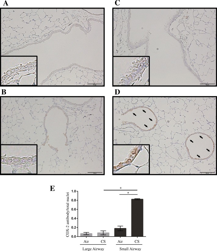
Effects of CS exposure on COX-2 expression in the lungs of C57BL/6 J mice. Mice were exposed to either 3.5% CS or fresh air (control) for 30 min/day for 5 days. Representative immunohistochemistry images of (a) the large-airway area in a control lung, (b) the small-airway area in a control lung, (c) the large-airway area in a CS-exposed lung, and (d) the small-airway area in a CS-exposed lung. Brown color indicates COX-2-positive airway epithelial cells (arrows). Scale bar, 100 μm. Insets are high magnification views of airway epithelial cells. e The ratio of COX-2-positive nuclei to the total count of nuclei present in a field at 400× magnification and determined in 10 different areas of the lung per mouse. Values are shown as the mean ± SEM (n = 3/group). *p < 0.001. CS, cigarette smoke; COX-2, cyclooxygenase-2

