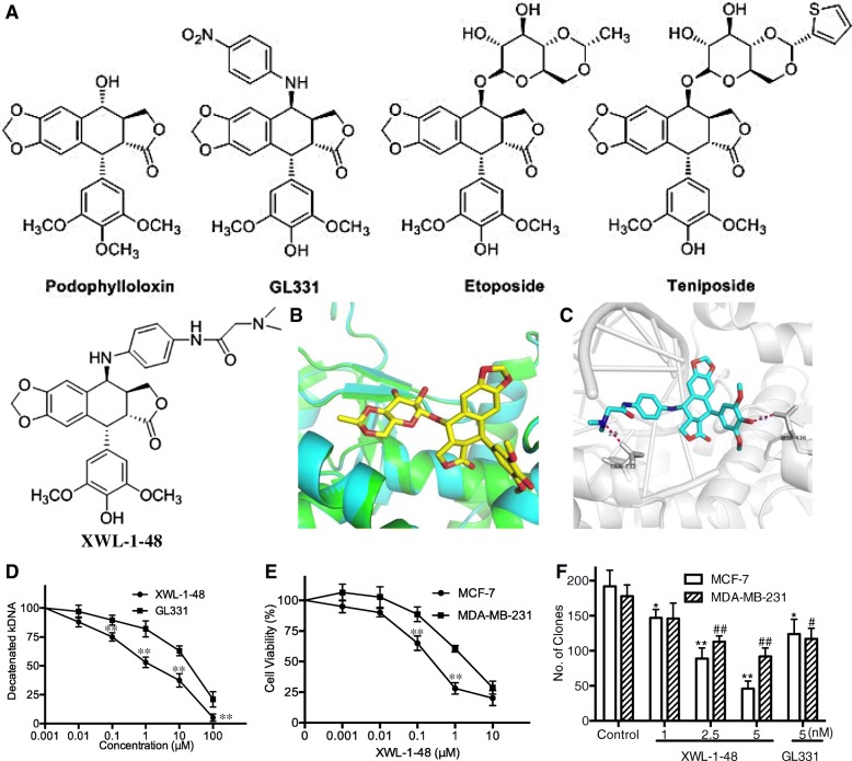
Fig. 1.
Effect of XWL-1-48 on TopoII activity and growth of breast cancer cells. a The chemical structure of podophylloloxin, GL331, Etoposide (VP16), Teniposide and XWL-1-48 is shown; b Superimposition of Top2α homology model (cyan) and Top2β crystal structure (Green), shown in cartoon representation. XWL-1-48 pose from the Top2β crystal structure (stick representation, carbons yellow) shown for clarity; c Molecular docking study of XWL-1-48 with carbons in cyan to crystal structure of Top2α-DNA homology model. Only interacting residues were labeled. Hydrogen bonds are shown as purple dotted lines; d To determine the effect of XWL-1-48 on TopoII activity, kDNA decatenation assay was performed as described in materials and methods; e The cytotoxic effect of XWL-1-48 on growth of MCF-7 and MDA-MB-231 cells was determined by MTT assay. IC50 was calculated by Prism6.0 software; f MCF-7 or MDA-MB-231 cells were seeded in a 6-well plate, and incubated with XWL-1-48 (1, 2.5, 5 nM) or GL331 (5 nM) for 24 h. After 10–14 days, colonies (> 50 cells) were fixed and manually counted. A representative of three experiments is shown. Data were shown as mean ± SD of three independent experiments. *p < 0.05, **p < 0.01 vs. control

