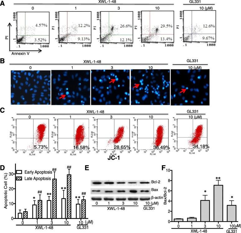
Fig. 4.
Effect of XWL-1-48 on cellular apoptosis of breast cancer. a, d Treatment with XWL-1-48 (1, 3, 10 μM) and GL331 (10 μM) for 24 h, early and late apoptotic cells were detected by flow cytometry. A representative of three independent experiments is shown; b Apoptosis of MCF-7 cells was detected by DAPI staining. MCF-7 cells were treated by XWL-1-48 or GL331 for 24 h, then the cells harvested, fixed and stained with 4, 6-diamidino-2-phenylindole (DAPI); c Loss mitochondrial membrane potential of cells treated by XWL-1-48 or GL331. MCF-7 cells treated with or without XWL-1-48 and GL331 for 24 h at indicated concentrations. The cells were harvested and washed, and then cells were incubated for 20 min in freshly prepared JC-1 solution at 37 °C. Spare dye was removed by dye buffer solution washing and cell-associated fluorescence was measured by flow cytometry; e, f MCF-7 cells were incubated with XWL-1-48 (1, 3, 10 μM) and GL331 (10 μM) for 24 h, expression of Bcl2, Bax were determined by western blot analysis. A representative result is shown from at least three independent experiments. Data were shown as mean ± SD of three independent experiments, *, p < 0.05, **, p < 0.01 or #, p < 0.05, ##, p < 0.01 vs. control group, respectively

