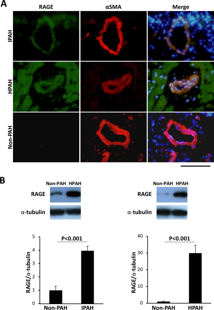
Fig 1. RAGE expression in PASMCs of patients with PAH.

A. Immunohistochemical staining of RAGE (green) (left figures), α-SMA (red) (center figures) and merge images (right figures) in distal pulmonary arteries of patients with IPAH (upper figures), patients with HPAH (middle figures) and non-PAH patients (lower figures). Nuclear staining was performed by using DAPI (blue). Bar = 100 μm. B. Western blot analysis of RAGE in PASMCs of patients without PAH (non-PAH) versus patients with IPAH (left) and patients without PAH (non-PAH) versus patients with HPAH (right). Data are mean ± SD.
