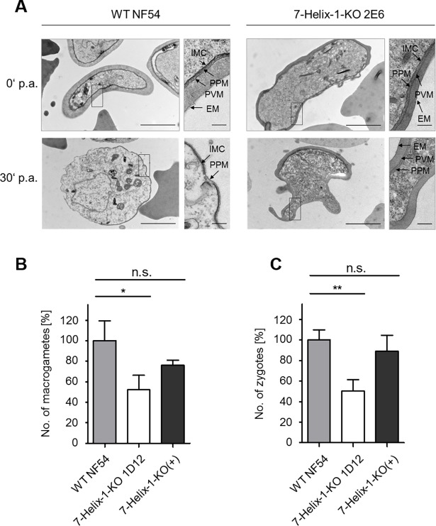
Fig 4. Ultrastructural analysis of activated 7-Helix-1-KO gametocytes and phenotype rescue.
(A) Ultrastructure of 7-Helix-1-KO gametocytes. Transmission electron microscopic analyses of WT NF54 and 7-Helix-1-KO 2E6 gametocytes at 0 and 30 min p.a.. EM, erythrocyte membrane; IMC, inner membrane complex; PPM, parasite plasma membrane; PVM, parasitophorous vacuole membrane. Bar, 2 μm; enlargement, 0.2 μm. (B, C) 7-Helix-1-KO phenotype rescue by episomal complementation. Gametocytes of WT NF54, 7-Helix-1-KO 1D12 and 7-Helix-1-KO(+) were activated in vitro. Samples were taken at 30 min (macrogametes, B) and 4 h (zygotes, C) p.a. and immunolabeled with anti-Pfs25 antibody. The numbers of parasites were counted in 30 optical fields in triplicate (mean ± SD). The numbers of WT parasites were set to 100%. n.s., not significant; * p ≤ 0.05; ** p ≤ 0.01 (One-Way ANOVA with Post-Hoc Bonferroni Multiple Comparison test). Results (in B and C) are representative of three independent experiments.

