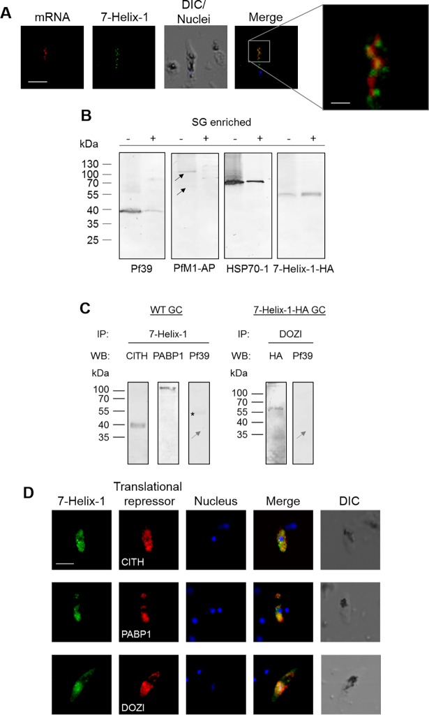
Fig 6. Localization of 7-Helix-1 in SGs and interaction with translational repressors.
(A) Co-localization of 7-Helix-1 with mRNA-aggregates. Mature WF NF54 gametocytes were subjected to mRNA-FISH-IFA and mRNA was labeled with a biotinylated oligo-dT25 probe (red); counterlabeling was performed using mouse anti-7-Helix-1rp2 antisera (green). Nuclei were highlighted by Hoechst33342 nuclear stain (blue). Frame indicates the area chosen for enlargement. DIC, differential interference contrast. Bar, 5 μm; enlargement, 1 μm. (B) Accumulation of 7-Helix-1 in SG fractions. Gametocytes of line 7-Helix-1-HA were stressed by treatment with sodium arsenite for 1 h and a SG core fraction enrichment was conducted. Lysates of 7-Helix-1-HA gametocytes (-) and of enriched SGs (+) were subjected to WB, using mouse antisera directed against Pf39 (~39 kDa) or PfM1-AP (~126 and 68 kDa, black arrows) or rabbit antibodies against HSP70-1 (~70 kDa) or the HA-tag to detect 7-Helix-1-HA (~60 kDa). (C) Co-immunoprecipitation of 7-Helix-1 with CITH, PABP1 and DOZI. Lysates of WT NF54 or 7-Helix-1-HA gametocytes were subjected to co-immunoprecipitation assays using polyclonal mouse anti-7-Helix-1rp2 antisera or polyclonal rabbit anti-DOZI antisera, followed by WB using rabbit anti-CITH, anti-PABP1 and anti-HA antibodies or mouse anti-Pf39 antibody to detect precipitated proteins. Grey arrow indicates the expected running line in the negative control. Asterisk indicates a band corresponding to the precipitation antibody. (D) Co-localization of 7-Helix-1 with CITH, PABP1 and DOZI. WT NF54 gametocytes were immunolabeled with mouse anti-7-Helix-1rp2 antisera (green) and rabbit anti-CITH, anti-PABP1, or anti-DOZI antibodies (red). Nuclei were highlighted by Hoechst33342 nuclear stain (blue). DIC, differential interference contrast. Bar, 5 μm. Results are representative of three independent experiments.

