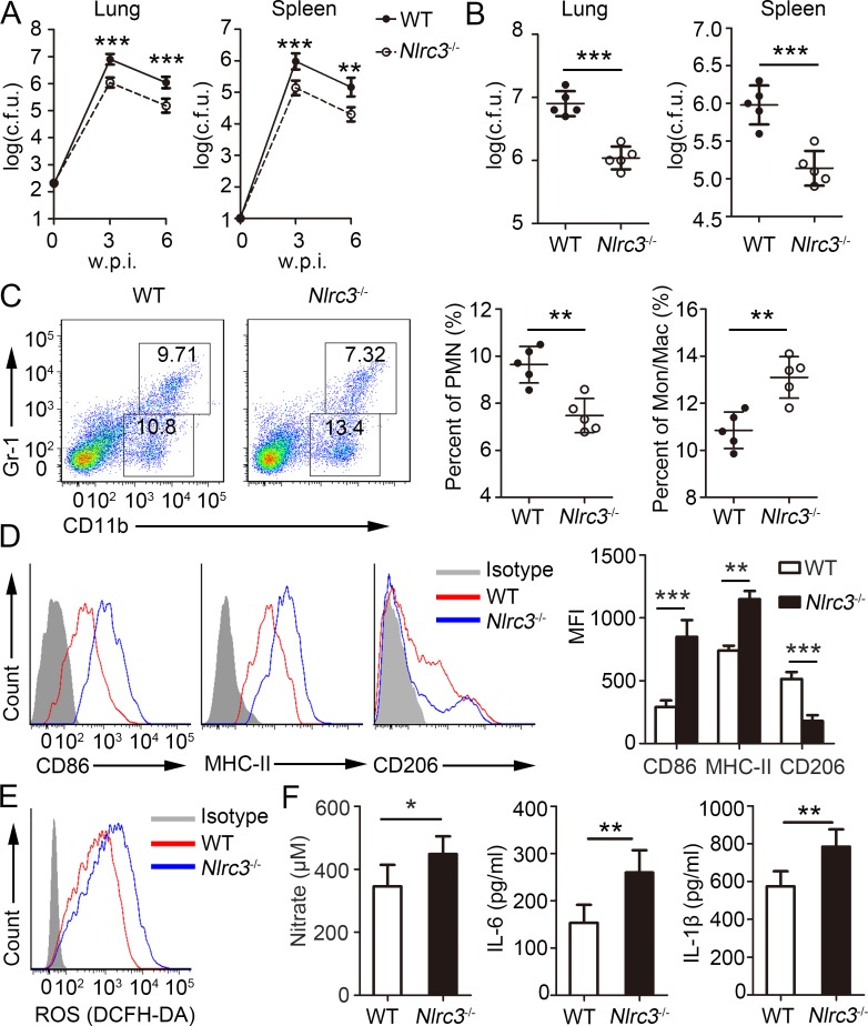
Fig 2. Nlrc3-/- mice are protected from M. tuberculosis infection.
WT and Nlrc3-/- mice infected with approximately 200 colony-forming units (c.f.u.) of M. tuberculosis were monitored. (A) Bacterial burdens were determined after infection at 3 and 6 w.p.i.. (B) Bacterial burdens were determined after infection at 3 w.p.i.. (C) Frequencies of lung-infiltrating cells that are neutrophils (CD11b+ Gr-1+) or monocyte-macrophages (CD11b+ Gr-1-) at 3 w.p.i.. (D) Expressions of CD86, MHC-II and CD206 were detected on monocyte-macrophages (CD11b+ Gr-1-) via flow cytometry at 3 w.p.i.. (E) ROS production by monocyte-macrophages (CD11b+ Gr-1-) were detected assessed as mean fluorescence intensity (MFI) of intracellular CFDA. (F) Concentrations of nitrate were measured by nitrate reductase assay and concentrations of IL-6 and IL-1β in lungs (homogenized in 2 ml PBS and 0.05% Tween 80) were detected by ELISA at 3 w.p.i.. Data shown in are the mean ±SD. *P < 0.05, **P < 0.01 and ***P < 0.001. Data are representative of three independent experiments with similar results.

