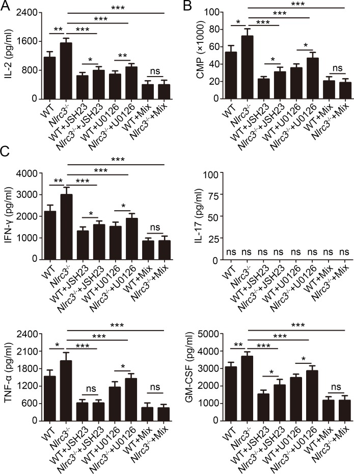
Fig 7. NLRC3 suppresses activation of CD4+ T cells via negatively regulating NF-κB and ERK Signaling.
Purified WT and Nlrc3-/- CD4+ T cells were stimulated for 48 hr with anti-CD3 (1 μg/ml) plus anti-CD28 (1 μg/ml) in the presence or absence of the NF-κB inhibitor JSH-23 (20 μM), MEK1/2-inhibitor U0126 (40 μM) or the mix of the two. (A) Concentrations of IL-2 in supernatants were detected by ELISA. (B) The incorporation of thymidine was measured during the final 8 hr. (C) Concentrations of IFN-γ, IL-17, TNF-α and GM-CSF in supernatants were detected by ELISA. Data shown are the mean ±SD. *P < 0.05, **P < 0.01 and ***P < 0.001. Data are representative of three independent experiments with similar results.

