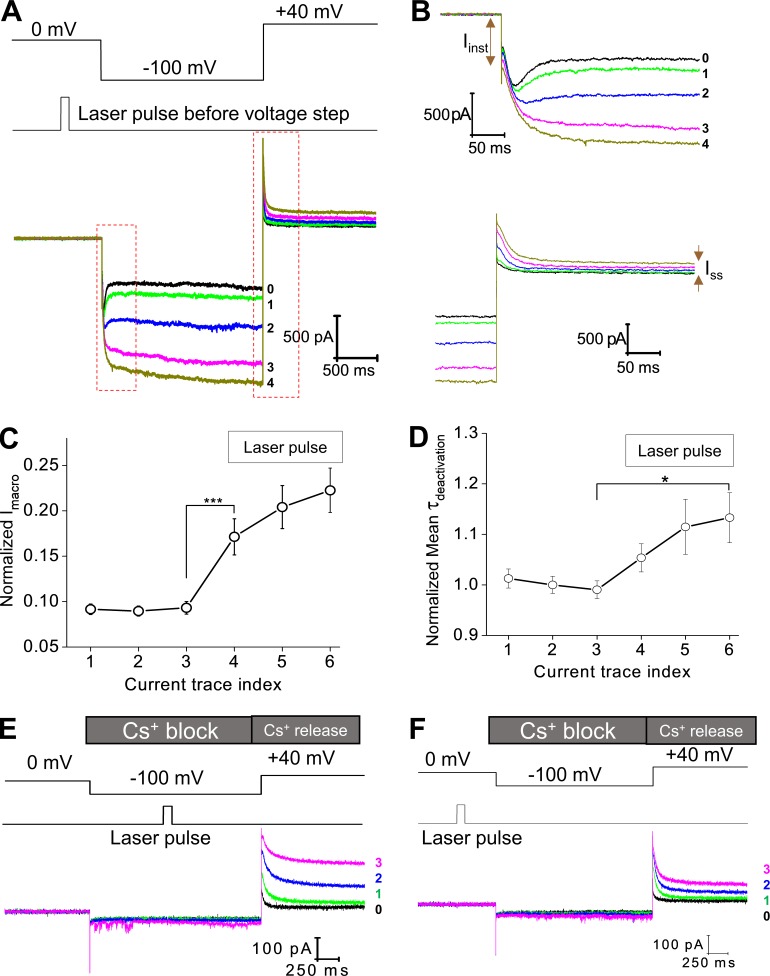
Figure 3.
Photodynamic modification of the spHCN channel in the closed state and the sensitivity of modified spHCN channels to Cs+. (A) Top: Voltage command and the timing of laser pulses. Laser pulses were applied preceding the hyperpolarization voltage step when most of the channels should stay in the closed state. Bottom: Current traces recorded with 1 µM FITC-cAMP in the bath solution. The last control trace before laser pulse is shown in black and labeled 0. (B) Zoomed views of the region within the red box shown in A. (C) Averaged (n = 15) results showing the effect of laser pulses on the amplitude of macroscopic current. ***, P ≤ 0.001. (D) Averaged (n = 15) results showing the effect of laser pulses on the time constant of deactivation. *, P ≤ 0.05. Error bars represent SEM. (E) During the −100-mV voltage step, Cs+ applied to the extracellular side of the membrane blocks the spHCN current after photodynamic modification (laser pulse during voltage step). The following depolarizing voltage step from −100 to +40 mV released the block by Cs+ and revealed the effects of photodynamic modification. Photodynamic modification leads to slowdown in channel deactivation and increases in Ih(tail) and Iss. Black, last current trace before laser pulse. Green, traces with laser pulses. (F) Laser pulses were applied preceding the hyperpolarization voltage step. Cs+ blocks the macroscopic current at −100 mV. The voltage step from −100 to +40 mV released the Cs+ block and revealed the effects of photodynamic modification. Detailed analyses are shown in Fig. S5.

