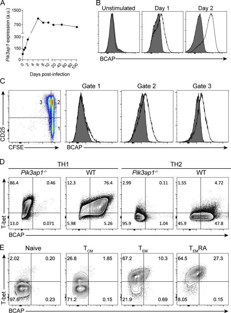
Figure 1.
BCAP is up-regulated in activated CD8+ T cells. (A) Expression of Pik3ap1 mRNA by splenic CD8+ OT-I T cells at the indicated times following infection with LM-OVA. Data are from the Immunological Genome Project. (B) Flow cytometry analysis of BCAP expression by CD8+ T cells from WT (open histograms) or Pik3ap1−/− (filled histograms) mice at the indicated times following stimulation with plate-bound anti-CD3/anti-CD28. Data are representative of more than five independent experiments. (C) Flow cytometry analysis of CFSE dilution and CD25 expression by WT CD8+ T cells activated for 29 h with plate-bound anti-CD3/anti-CD28 + IL-2 (left) and BCAP expression by WT (open histograms) or Pik3ap1−/− (filled histograms) cells in each of the indicated gates. (D) Flow cytometry analysis of BCAP and T-bet expression by WT or Pik3ap1−/− CD4+ T cells activated and polarized under TH1 or TH2 conditions as indicated. (E) Flow cytometry analysis of BCAP and T-bet expression by gated naive, TCM, TEM, and TEMRA CD8+ T cells from human peripheral blood as indicated. (C–E) Data are representative of three independent experiments.

