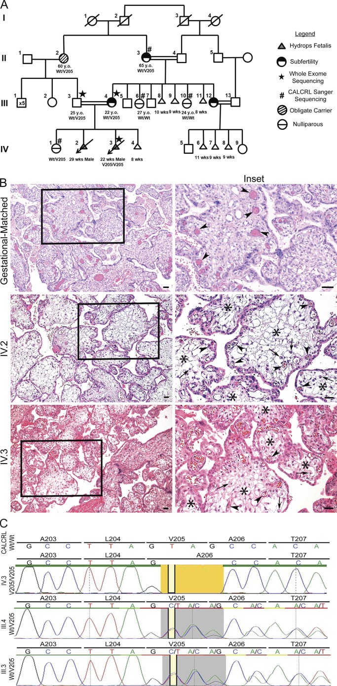
Figure 1.
Familial pedigree, placental histology and sequencing traces of family with NIHF. (A) Whole exome sequencing identified mutations in subject IV.3 in hCALCRL following the elimination of all previously implicated candidate genes. Parents, III.3 and III.4, and maternal grandmother were confirmed as heterozygous carriers of the hCALCRL(V205del) variant. Family history gives rise to two phenotypes dependent on haplotype: subfertility and nonimmune HF. (B) Placental histology from affected homozygous fetuses (IV.2 and IV.3) compared with a gestational-matched, normal placenta. The right column represents enlargements of boxed areas in the left column. Arrowheads indicate fetal vessels. In the case of the normal placenta, these vessels are completely filled with fetal erythrocytes and distributed both at the periphery and closer to the central villus core. In the case of the two affected placentas, the fetal vessels are more commonly found near the villus periphery, are compressed, and contain fewer erythrocytes. Arrows indicate presence of scattered Hofbauer cells. Asterisks indicate regions of severe edema within the chorionic villi of the affected placentas, a finding not seen in the normal placenta. (C) Whole exome sequencing traces for IV.3, III.4, and III.3 showing the amino acid residue valine 205 deletion (yellow). In individuals III.4 and III.3, the heterozygous allele (gray) is represented by the double peak.

