Figure 1.
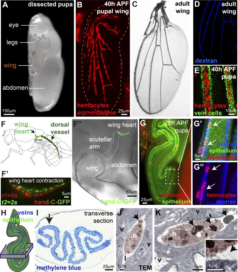
Drosophila pupal wing veins carry circulating hemocytes. (A) 75-h APF Drosophila pupa dissected from pupal case for imaging. (B–D) Hemocytes (red, srp>moesin-mCherry, B) restricted to wing veins by 40 h APF (B) in a pattern reminiscent of adult vessels (C, brightfield, and D, dextran-loaded veins, blue). (E) Hemocytes (red) within lumens of 40-h APF wing veins (vein wall cells labeled using shortvein>GFP, green). (F) Wing hearts (F) contract (F’ and F’’, hand-C-GFP reporter) to pump hemolymph into the veins. (G) 75-h APF wings are folded (ubiquitous Moesin-GFP, green), but hemocytes (arrows, red, srp>mch-moesin) remain within vein lumens (G’ and G’’, blue dextran). (H and I) Folded nature of wing revealed by resin histology (H, schematic, and I, methylene blue; arrow in I indicates flat epithelium adjacent to vein). (J and K) Hemocytes (h, false-colored pink in transmission electron micrograph, TEM) within the lumen (labeled l) of 75-h APF wing veins (v, vein wall cells, false-colored blue) contain large cytoplasmic granules (arrows, K) and are occasionally tethered to the vein wall (arrow, inset K). Also see Fig. S1 and Videos 1, 2, 3, 4, and 5.

