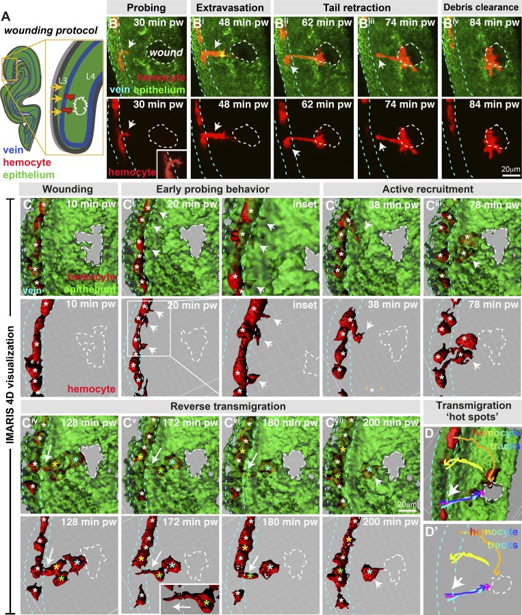
Figure 2.
Drosophila wing vein hemocytes extravasate from vessels to wounds. (A and B) Wounding of pupal wing epithelium (green, ubiquitous GFP-Moesin; schematic, A, and in vivo imaging, B) triggers hemocyte (red, srp>mch-moesin) extravasation from wing veins (blue dashed lines, position determined from z-sections) to sites of damage (white dashed lines). (C) IMARIS software permits 3D visualization of imaging data. Hemocytes (red) probe the vessel wall (arrows, B and insets in B and Ci) and extravasate from the vessel (arrows, Bi–Biii and Cii), requiring retraction of the hemocyte tail (arrow, Biii). Multiple hemocytes exit the vessel (Civ), but some return to the vessel (arrows, Civ–Cvii; individual hemocytes indicated by asterisks: white, all hemocytes in C–Ciii and hemocytes in vessels, Civ–Cvii; cyan, extravasated hemocytes that remain at the wound; yellow and green, reverse migrating hemocytes). (D) Tracking hemocyte trajectories indicates multiple hemocytes extravasating from similar vein wall locations (arrows; blue, cyan, and magenta tracks), although hemocytes also exit from additional locations (yellow and orange tracks). pw, post-wounding. Also see Videos 6 and 7.

