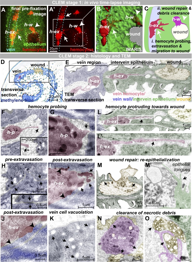
Figure 3.
CLEM of hemocyte extravasation. (A) In vivo imaging of extravasation (hemocytes, red, srp>mch-moesin; and epithelium, green, ubiquitous GFP-Moesin). (A–C) Samples fixed at key moments (hemocyte, h-ex, initiating transmigration; and hemocyte, h-w, reached the wound). Arrows indicate hemocytes in the process of extravasation (h-ex), and arrowheads indicate extravasated hemocytes at wound (h-w). (D–O) Samples prepared for TEM. (D and E) Resin transverse section (D, methylene blue) and TEM section (E) illustrate the route from vein to wound with a hemocyte (h-ex, false-colored red) traversing the vein wall (E). (F and G) TEM reveals hemocyte probing of the vessel wall, with long (F) and short (G) protrusions targeting junctions between adjacent vein wall cells (arrow, G). Arrows (black, F) indicate the end of the long hemocyte probing protrusion. White arrow (F, inset) indicates intact vein wall junction. (H–K) Prior to extravasation, the vein wall barrier is intact (H; arrows, vein wall junctions), but extravasating hemocytes move across and flatten this vein wall barrier (arrowheads, I and J). Extravasating hemocytes are associated with large darkly staining deposits (arrows, I), and vein cells exhibit vacuolation (arrows, K). (L–O) Hemocytes use long protrusions to migrate through the extravascular space toward the wound (arrow, L). Wounds are repaired by long epithelial tongues (arrows, M and M'). Hemocytes at the wound site (magenta, N and O) clear up necrotic wound debris (yellow, indicated by arrows). All images were taken from two 75-h APF Drosophila pupae prepared for CLEM. Also see Fig. S2.

