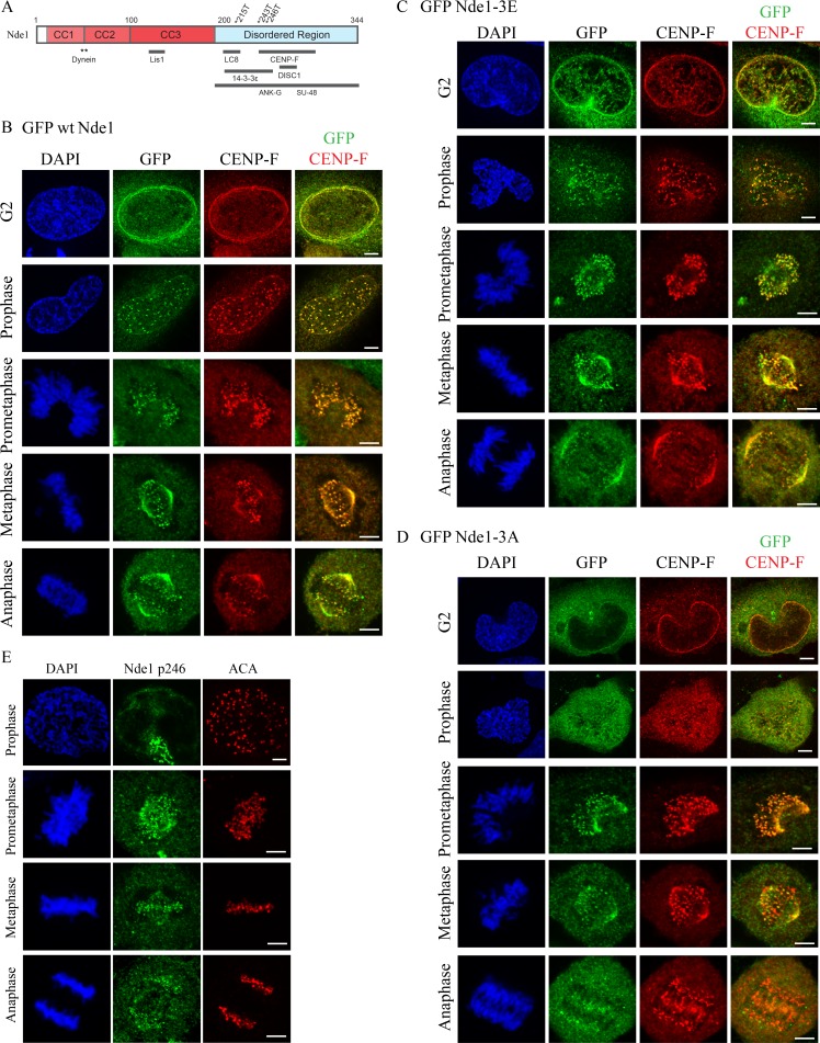
Figure 1.
Subcellular localization of Cdk1 phosphomimetic and phosphomutant Nde1 in G2 and mitosis. (A) Diagram of Nde1 showing Cdk1 phosphorylation sites (T215, T243, and T246) and interaction sites. (B–D) HeLa cells were transfected with GFP-tagged WT, phosphomimetic, and phosphomutant Nde1 and examined for localization to the G2 NE and mitotic kinetochores. GFP was detected by immunocytochemistry. WT and phospho-Nde1 colocalize with CENP-F at the NE and prophase–anaphase kinetochores. The phosphomutant Nde1 was weakly detected at these sites and absent from metaphase and anaphase kinetochores. (E) HeLa cells were stained with CDK1-phospho-specific antibody (p246; Alkuraya et al., 2011), which reacted with prometaphase-to-anaphase kinetochores, consistent with the distribution of the expressed phosphomimetic Nde1. Bars, 5 µm.

