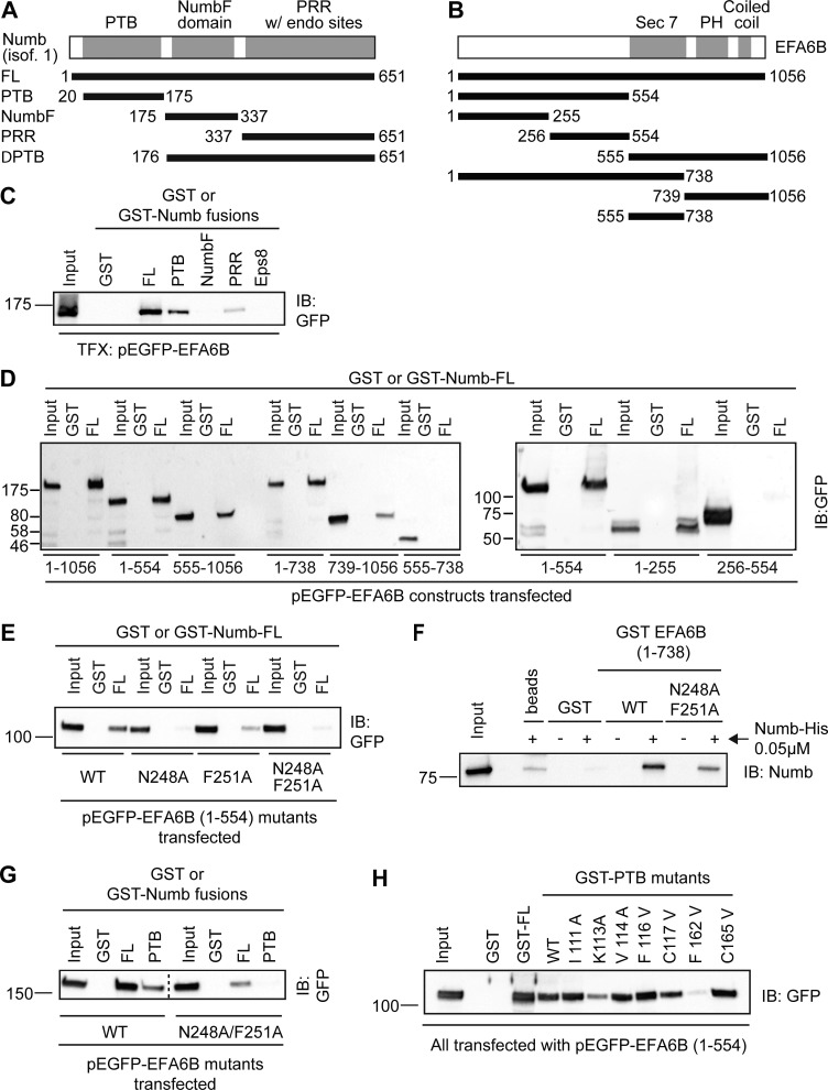
Figure 6.
The N-terminal NPLF motif of EFA6B is required for binding to NUMB via its PTB domain. (A and B) Scheme of NUMB (A) and EFA6B (B) domain organization and of the fragments used to characterize the interaction between the two proteins. Numbers correspond to amino acid boundaries. (C) Lysates (2.5 mg) of Phoenix cells transfected with pEGFP-EFA6B were incubated with 0.5 μM GST or GST-fused to NUMB of the indicated NUMB fragments (PTB, 20–175 aa; NUMBF, 175–337 aa; NUMB-PRR, 337–651 aa) or GST-PTB of EPS8, used as negative control. Input lysates (1%) and bound material were analyzed by IB with anti-GFP to visualize EFA6B or by Ponceau staining to detect recombinant GST-fusion proteins (shown in Fig. S3 A). MW markers are shown on the left in kilodaltons. TFX, transfected. (D) Lysates (1.5 mg) of Phoenix cells transfected with either the full-length pEGFP-EFA6B or the indicated GFP-fused EFA6B fragments (amino acid boundaries indicated at the bottom) were incubated with 0.5 μM GST or GST-NUMB immobilized on beads. Input lysates (1% of the total) and bound material were analyzed by IB with anti-GFP to visualize EFA6B and its deletion mutants, or by Ponceau staining to detect the recombinant GST proteins (see Fig. S3 C). MW markers are shown on the left in kilodaltons. (E) Lysates (750 μg) of Phoenix cells transfected with pEGFP-EFA6B 1–554 either WT or carrying N248A, F251A point mutations, alone or in combination, were incubated with 0.5 μM GST or GST-NUMB immobilized on beads. Input lysates (2%) and bound material were analyzed by IB with anti-GFP to visualize EFA6B and its point mutants, or by Ponceau staining to detect recombinant GST proteins (see Fig. S3 D). MW markers are on the left in kilodaltons. (F) Purified, His-tagged His-NUMB (0.05 μM) was incubated with 0.5 μM GST-EFA6B 1–738 either WT or carrying double N248A/F251A point mutations, or with GST alone (control) or in the absence of any protein (beads) to rule out unspecific signals of the antibodies. Input (1/5 of total) and bound material were analyzed by IB with anti-NUMB to visualize NUMB. The Ponceau staining to detect GST recombinant proteins is in Fig. S3 F. (G) Lysates (2 mg) of Phoenix cells transfected with pEGFP-EFA6B WT or pEGFP-EFA6B N248A-F251A were incubated with 0.5 μM GST, GST-NUMB, or GST-PTB immobilized on beads. Input lysates (2%) and bound material were analyzed by IB with anti-GFP to visualize EFA6B. Ponceau staining to detect the recombinant GST proteins is in Fig. S3 H. Dashed line indicates that the intervening lanes have been spliced out. (H) Lysates (2 mg) of Phoenix cells transfected with pEGFP-EFA6B 1–554 were incubated with 0.5 μM GST, GST-NUMB, GST-PTB, or the indicated GST-PTB point mutants immobilized on beads. Input lysates (2%) and bound material were analyzed by IB with anti-GFP to visualize EFA6B. Ponceau staining to detect the recombinant GST proteins is in Fig. S3 J. (F–H) MW markers are shown on the left in kilodaltons.

