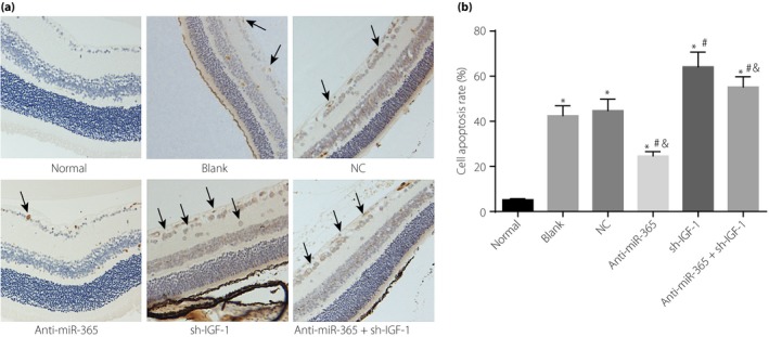
Figure 9.

Apoptosis rate of retinal neurons in retinal tissues among six groups. (a) Terminal deoxynucleotidyl transferase dUTP nick‐end labeling staining of retinal neurons in retinal tissues of six groups. (b) The apoptosis rate of retinal neurons in retinal tissues among six groups; the cells shown by black arrows were apoptotic retinal neurons. *P < 0.05, compared with the normal group; # P < 0.05, compared with the blank group; & P < 0.05, compared with the transfected insulin‐like growth factor (IGF‐1) short hairpin ribonucleic acid plasmid (sh‐IGF‐1) group. miR‐365, micro‐ribonucleic acid‐365; NC, negative control.
