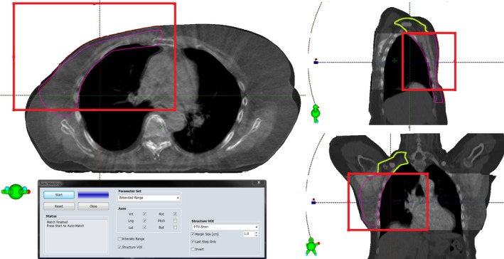
Figure 1.

Region of interest for matching the CBCT image to the CT image is drawn with the red rectangle. The matching region of interest is centered to the PTV (red contour), effectively excluding the spine. The division of PTV‐5 mm into PTVb/c (magneta contour) and PTVsclav (green contour) is made on the level where the original PTV (red contour) reaches the skin, shown in the sagittal and coronal views.
