
Fig. 2. CRSBP-1 ligands stimulate contraction of CRSBP-1-associated fibrillar structures in SVEC4–10 cells.
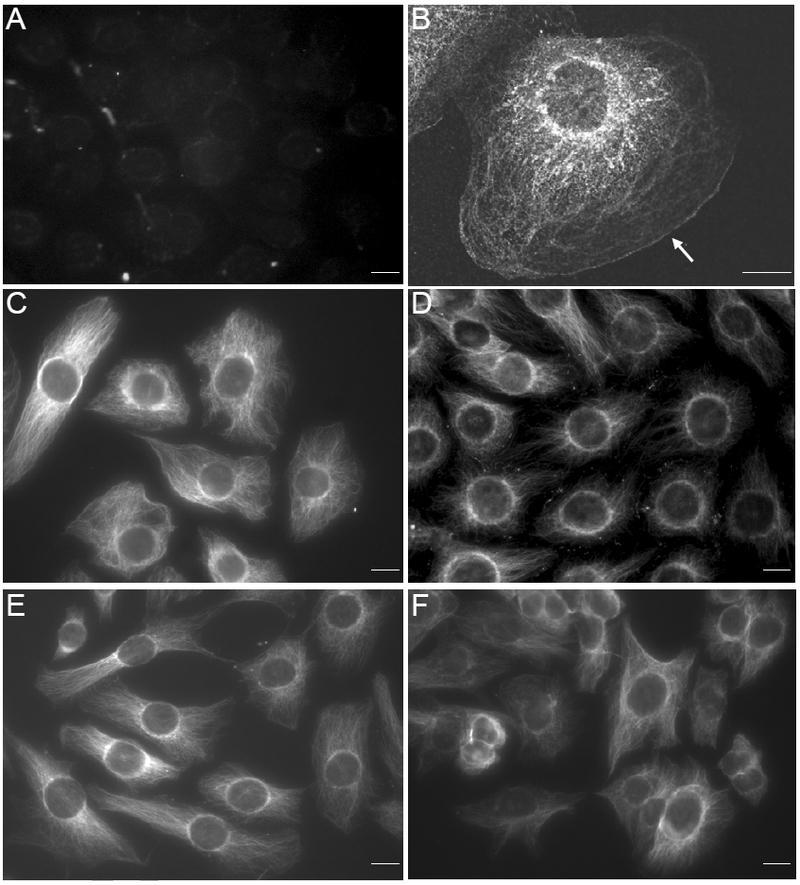
Cells were seeded on Petri dishes (panel B) and cover slips (panels A, C, D, E and F). Cells were treated with vehicle only (panels A, B and C), VEGF-A peptide (10 μM) (panel D), HA (100 μg/ml) (panel E) or HA (100 μg/ml) plus VEGF-A pep tid e (1 0 μM) (panel F). After 2 h at 37°C, cells were fixed with paraformaldehyde and then permeabilized by acetone (panel B) or fixed and permeabilized with methanol at −20°C for 10 min (panels A, C, D, E and F). CRSBP-1 was stained with anti-CRSBP-1 antiserum (panels B, C, D, E and F) or preimmune serum panel (panel A) followed by FITC-conjugated goat anti-rabbit IgG and visualized with a fluorescence microscope. The bar indicates a scale of 10 μm (panel A). The arrow in (panel B) indicates the edge of the plasma membrane. Control staining (with preimmune serum) of cells treated with CRSBP-1 ligands were negative like (panel A) (data not shown). Data are representative of five independent experiments.