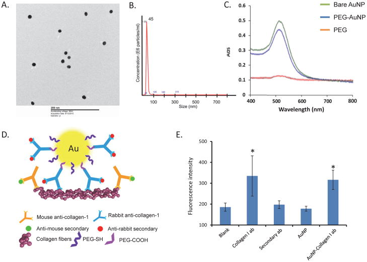
Figure 1.
A. Electron microscope image showing AuNPs of approximately 20nm in size. B. PEG-coated AuNPs measured 45nm in size with nanoparticle tracking analysis. C. UV-Vis spectroscopy showing that peak absorbance of both bare and PEG-coated AuNPs at 525nm. D. A schematic of the experimental approach for staining. An anti-rabbit secondary antibody was used to probe collagen-I-AuNPs collagen in plate or kidney tissue, while mouse collagen I and anti-mouse secondary antibody served as positive controls in kidney tissue. E. Co-I-AuNPs reached signal similar to the same concentration of collagen-I antibody on a collagen coated plate. *p<0.05 vs. Blank

