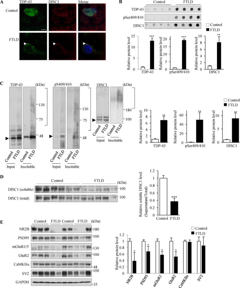
Figure 5.

TDP-43 aggregates sequestered endogenous DISC1 in FTLD patient brain. (A) DISC1 and TDP-43 are mislocalized and co-aggregated in the cytosol in FTLD patient brains. DISC1 and TDP-43 were immunostained using anti-DISC1 antibody (h598C) and anti-TDP43 antibodies, respectively, in control (Control, top panels) and FTLD patient brains (FTLD, bottom panels). Brain samples from three different FTLD patients were stained. In neurons containing TDP-43 aggregates, 89% of TDP-43 aggregates were DISC1-positive (n=27). Other images immunostained with another anti-DISC1 antibody (HM6-5) are shown in Supplemental Figure S8D. Scale bar represents 5 μm. (B–D) Insoluble DISC1 levels were increased while soluble DISC1 levels were decreased in FTLD patient brains. (B) 125 μg of the homogenates of temporal lobes from control (n=5) and FTLD (n=5) subjects were incubated with 2% sarkosyl and amounts of insoluble proteins were analyzed by the filter-trap assay, followed by immunoblotting with indicated antibodies (h598C for DISC1). The levels of each genes relative to those in control are also shown (below) (TDP43: t=7.833, ***P<0.0001; pSer409/410: t=12.928, ***P<0.0001; DISC1: t=4.051, **P=0.0037, unpaired two-tailed t-tes). (C) The homogenates of temporal lobe from control (n=6) and FTLD (n=5) subjects were incubated with 2% sarkosyl and then centrifuged at 100,000×g. The resulting sarkosyl-insoluble pellets and the input were analyzed by western blotting with indicated antibodies (h598C for DISC1). Arrowheads indicate the band position of each monomeric protein. The levels of each genes relative to those in control are shown (right) (TDP43: t=3.246, **P=0.0098; pSer409/410: t=3.415, **P=0.0077; DISC1: t=3.253, **P=0.0099, unpaired two-tailed t-test). (D) The homogenates of temporal lobe from control (n=6) and FTLD (n=5) subjects were incubated with 2% sarkosyl and then centrifuged at 100,000×g. The resulting sarkosyl-soluble supernatans and the input were analyzed by western blotting with an anti DISC1 antibody (h598). The signal intensities of DISC1 in supernatants were once normalized to those in the input and then expressed as the relative ratio of control brains (right). (t=5.798, ***P=0.0003, unpaired two-tailed t-test). (E) Homogenates of temporal lobes from control (n=5) and FTLD patient brains (n=5) were subjected to western blotting with indicated antibodies (left). GAPDH was used as a loading control. The signal intensities of each gene relative to those in control brains are shown (right) (NR2B: t=2.689, *P=0.0275; PSD95: t=2.404, *P=0.0429; mGlulR1: t=2.369, *P=0.0453; GluR2: t=2.964, *P=0.0181, unpaired two-tailed t-test). Throughout the figures, error bars represent S.E.M.
