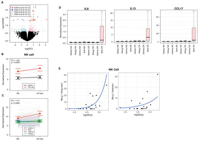
Fig. 4. NK cell gene activation is specific to allergic asthma.
(A) The Late group demonstrated significant differences in expression of similar genes in response to CR stimulation compared to controls (31 genes at FDR<0.05, 326 genes at FDR<0.25). (Stimulation linear model with fixed effects for site, gender, blood draw year, home allergen levels, and stimulation, and random effect for individual; nLate = 7, nNeither = 14). (B) The NK cell gene set showed significantly higher baseline expression in Late compared to Neither, and expression increased significantly with CR stimulation in Late (p-value and FC represent group term controlling for stimulation; number of individuals in each comparison is represented in parentheses; bounds represent 95% confidence intervals) (C) The NK cell gene set shows significantly higher baseline expression in all cases (Red; “Both” = Early and Late groups, n=20) compared to Neither (Black, n=102), asthma only (Blue; “Asthma”, n=40), and CR allergy only (Green; “Allergy”, n=35) groups and increases with CR stimulation only in the Both group. (D) Boxplot of normalized expression of IL9, IL13, and CCL17 showing group difference with allergen stimulation (Pink=CR-Early, Blue=DM-Early, Grey=Neither). (E) The average expression value of the NK cell gene set showed correlation with Bla g 1 levels measured in the living room (p<0.05) and bedroom (p=0.08) in the Both group.

