FIGURE 5.
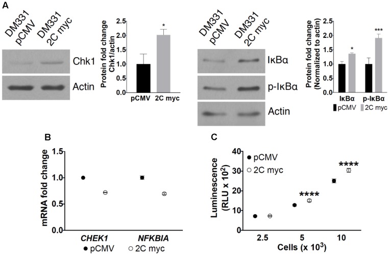
Effect of LAMP-2C expression on CMA substrates. (A) Cellular levels of CMA substrates Chk1, IκBα, and p-IκBα in DM331 pCMV and DM331 2C myc cells were examined by western blotting. Relative protein levels were calculated by setting the normalized expression to one for DM331 pCMV cells. (B) mRNA levels of CHEK1 and NFKBIA transcripts were analyzed by qPCR and normalized to ACTB expression. mRNA levels in DM331 pCMV cells were normalized and set to one. (C) Proteasome activity was measured using the Proteasome-Glo Chymotrypsin-Like Cell-Based Assay. Cells were incubated with a substrate succinyl-LLVY-aminoluciferin which penetrates into the cytoplasm. This substrate is cleaved by the proteasome to release aminoluciferin which is released from cells. Luciferase is added to these cells, cleaving aminoluciferin to a luminescent product detectable using a luminometer. Data were analyzed by two-way ANOVA or by two-tailed, unpaired Student’s t-test. ∗p < 0.05, ∗∗∗p < 0.001, and ∗∗∗∗p < 0.0001 (n = 3).

