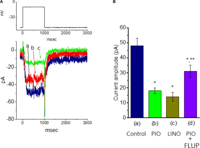
FIGURE 7.
Effect of PIO on IK(M) amplitude in mHippoE-14 hippocampal neurons. These experiments were conducted in cells bathed in high K+, Ca2+-free solution and the recording pipette was filled with K+-containing solution. (A) Superimposed IK(M) traces obtained in the absence (a) and presence of 3 μM (b), and 10 μM PIO (c). The upper part indicates the voltage protocol used. (B) Bar graph showing the effect of PIO, linopirdine, and PIO plus flupirtine on IK(M) amplitude (mean ± SEM; n = 9–11 for each bar). The IK(M) amplitude elicited by membrane depolarization from –50 to –10 mV was measured. (a) Control; (b) 10 μM PIO; (c) 10 μM linopirdine; (d) 10 μM PIO plus 10 μM flupirtine. ∗Significantly different from control (P < 0.05) and ∗∗significantly different from PIO (10 μM) alone group (P < 0.05). LINO, linopirdine; FLUP, flupirtine.

