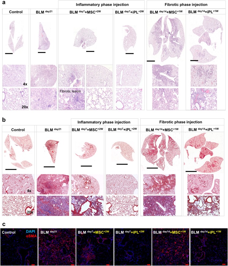
Fig. 6.
Treatment with AEC-II-iPL cells ameliorates severe pulmonary fibrosis. Representative images of lung histopathology are shown for all the experimental groups after 21 days of BLM-induced pulmonary fibrosis, the saline control, BLM untreated (BLM day21), and BLM cell-treated at day 7 (BLM day7 + iPL+2W, BLM day7 + MSC+2W) or day 14 (BLM day14 + iPL+1W, BLM day14 + MSC+1W). a lung sections were stained with hematoxylin–eosin, b Masson’s trichrome staining of all the experimental lungs showing a remarkable decrease of interstitial collagen deposition (the blue stain) in the BLM-injured lungs treated with AEC-II-iPL cells compared with the ones treated with MSC. c Immunostaining of lung sections of all experimental groups with nuclear stain DAPI (blue) and αSMA (red). Scale bar, 3 mm (a—whole section tile scan), 4 mm (b—whole section tile scan); 600 µm (a, b—×4); 200 µm (a, b—×20) and 100 µm (c)

