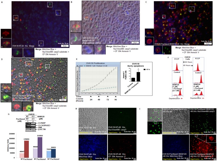
Figure 2.
Live Triple-fluorescence in OVK18 cells treated with paclitaxel plus BKM120. Live OVK18 cells, control and treated were stained with MitoView Blue + NucView488 casp3 substrate +CF 594 Annexin V (A–D). Photomicrographs were taken from live cells in culture within a span of 10 minutes. In separate experiments, the validation of the effect of paclitaxel (2.5 nM) plus BKM120 (1 µM) was tested by real-time proliferation (E, left panel), apoptosis (E, right panel), the mitochondrial potential (F), and expression of apoptotic markers by WB (G). The expression of apoptotic markers by WB was semi-quantified using Image-J and presented in a bar diagram with arbitrary units. The cleaved caspase3 activity, annexin V positivity and mitochondrial status of non-treated (H) and treated (I) live OKK18 cells at 48 hours are presented. A representative of uncropped blot representing the expression of proteins (cleaved PARP1, cleaved caspase3, BIM, and beta-actin) by WB from an independent experiment is presented in the Supplementary Figure.

