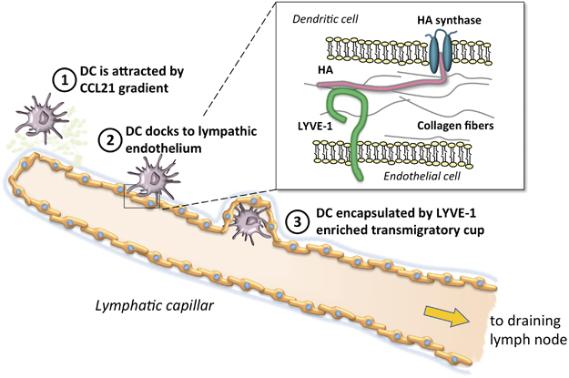
Figure 1.

Schematic representation of the major steps in DC migration to the lymphatic system. (1) Myeloid DCs in inflamed tissues are attracted to the regional lymphatic system in response to a CCL21 gradient (light green spots), released by endothelial cells, and move through gaps and portals in the basement membrane (light blue/gray); (2) DCs employ hyaluronan (HA) to anchor themselves to the basolateral surface of lymphatic endothelial cells; (3) DCs are enveloped by LYVE-1+ transmigratory cups to complete their migration into lymphatic capillary to reach draining lymph nodes. (Inset) HA (in pink) is synthesized at the inner face of the DC plasma membrane by hyaluronan synthases (in blue), and the growing HA polymer is extruded through the membrane to the outer surface. Endothelial cells express the LYVE-1 (in green) surface receptor for HA.
