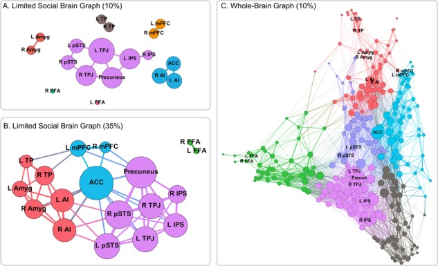
Fig. 4.

Network graphs. (A) When thresholding the 18-node social brain graph at a strict threshold, the graph is fractured with only a temporo-parietal and a salience cluster linking disparate regions. (B) However, at a more liberal threshold, there appear to be three large clusters: a temporo-parietal cluster (purple), a temporo-limbic cluster (orange) and a midline frontal cluster (green) linking them. (C) Within the context of the larger, whole-brain network, regions of the social brain are distributed throughout network space. Some regions such as the temporo-parietal regions (purple) show strong, centralized connectivity patterns, whereas anterior temporal and amygdala regions (orange) are located on the margins of the network.
