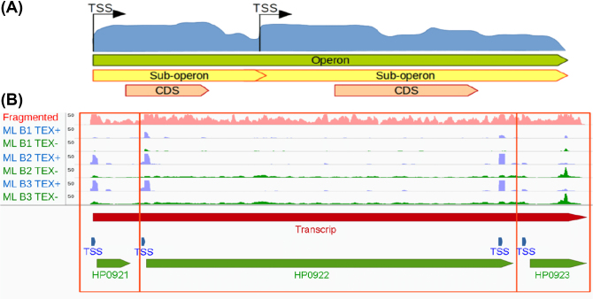
Figure 4:
Operon and sub-operon detection. (A) If there is more than one TSSs that does not overlap with genes located within one operon, the operon can be divided to several sub-operons based on these TSSs. (B) An example from H. pylori 26695. The coverage of RNA-seq with fragmentation, TEX+, and TEX- of dRNA-seq are shown in pink, blue, and green coverages, respectively. TSSs, transcripts/operons, and genes are presented as blue, red, and green bars, respectively. The two genes are located in the same operon but also in different sub-operons (two empty red squares).

