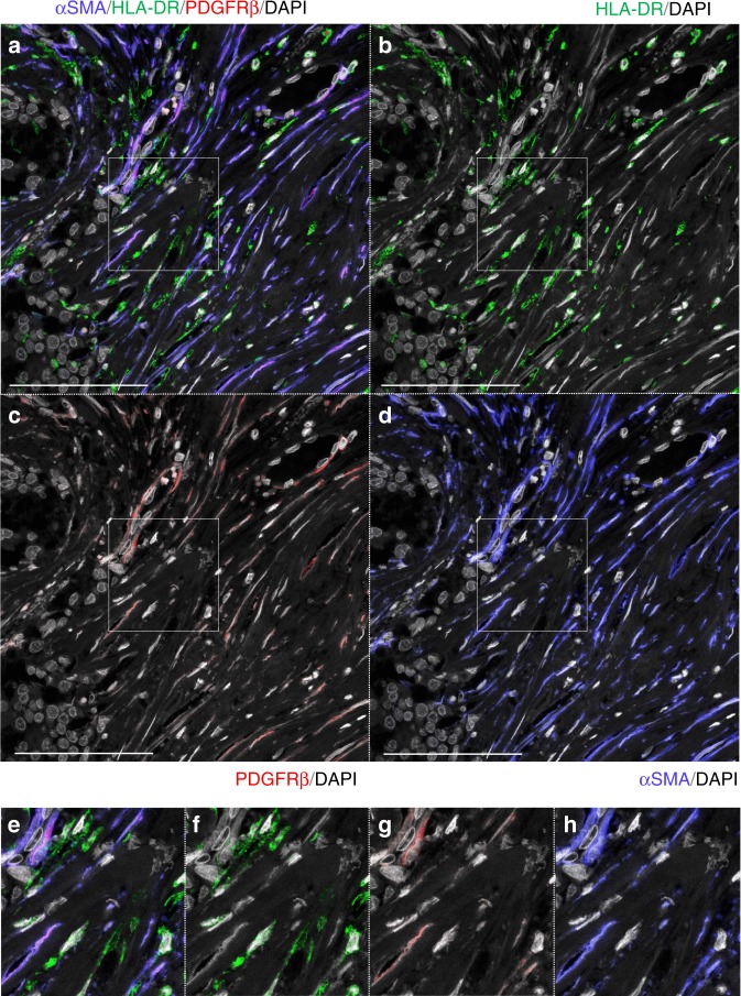
Fig. 4.
Triple immunostaining using markers of bone marrow stromal cell in case 1. αSMA, blue; PDGFRβ, red; HLA-DR, green; DAPI, gray. a–d Triple immunostaining with αSMA, HLA-DR, and PDGFRβ. αSMA(+)SCSSNs were almost identical to PDGFRβ(+)SCSSN (mean 99.3% (SD 0.6%) of αSMA(+)SCSSNs). and αSMA(+)HLA-DR(+) SCSSNs were sparse (mean 0.9% (SD 0.7%) of αSMA(+)SCSSNs). The analysis was carried out in six regions that consist of 5 HPFs (total 722 αSMA(+)SCSSNs). Scale bar: 100 μm. e–h Magnified images of the insets in a–d

