Fig. 8.
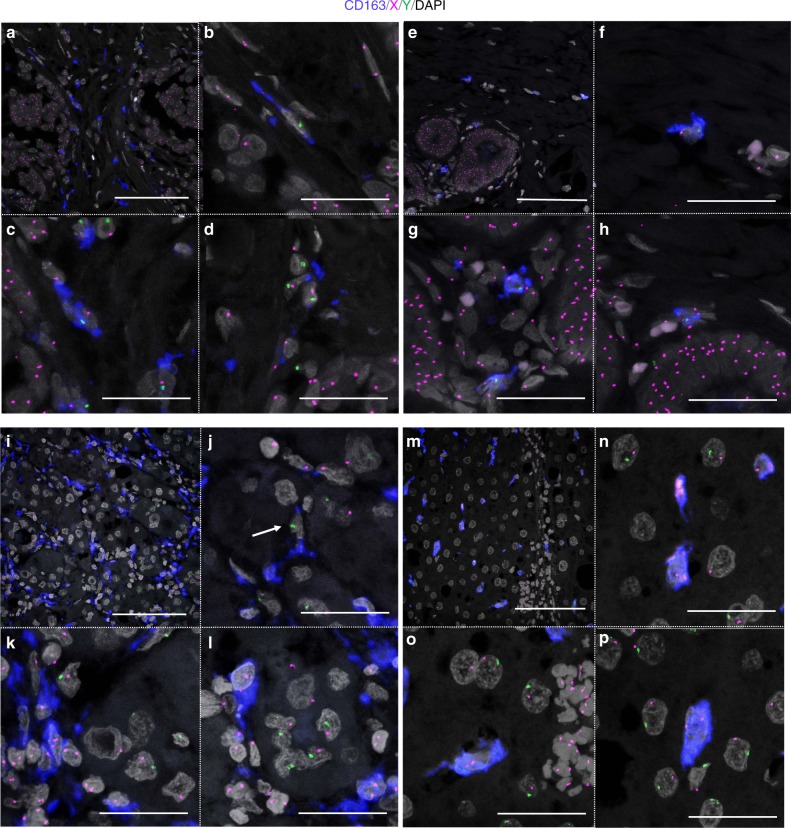
CD163-immunoFISH analysis in tumor and non-tumor tissues in cases 1 and 2. CD163, blue; X, magenta; Y, green; DAPI, gray. a–h Breast carcinoma in a 37-year-old female. a–d Tumor area. b–d Magnified images of a. e–h Non-tumor area. f–h Magnified images of e. Scale bars, a, e 100 μm and b–d, f–h 50 μm. i–p Hepatocellular carcinoma in a 64-year-old male. i–l Tumor area. j–l Magnified images of i. j The cell indicated by white arrow looks like a recipient-derived macrophage. However, when examined in the z-stack, recipient-derived CD163(−) cell and CD163(+) macrophages without the sex chromosome due to cutting artifact were overlapped in the z direction. m–p Non-tumor area. n–p Magnified images of m. Scale bars: i, m 100 μm and j–l, n–p 50 μm

