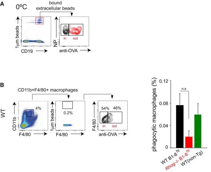
Figure EV3. Antigen‐specific splenic B cells phagocytose antigens in vivo through a BCR‐driven process also dependent on RhoG.

- WT B1‐8hi B cells were incubated in vitro with fluorescent 1 μm beads coated with NIP‐OVA at 0°C for 1 h and subsequently left unstained or stained with an anti‐OVA antibody. B cells with extracellular membrane‐attached beads that are not internalized are positive for the fluorescent beads and the anti‐ova staining (red histogram), while the control without primary antibody is in grey.
- Phagocytosis of 1 μm fluorescent beads covalently bound to NIP‐OVA by splenic macrophages from WT or Rhog −/− B1‐8hi and WT non‐transgenic mice was assessed after 5 h post‐IP immunization using extracellular staining with an anti‐ovalbumin antibody. The cytometry plot shows staining with the anti‐OVA antibody in the CD11b+ F4/80+ macrophage population. The graph on the right shows the percentage of phagocytic splenic macrophages in WT and Rhog −/− mice. Data represents means and SEM (n = 3). n.s, not significant (unpaired Student's t test).
