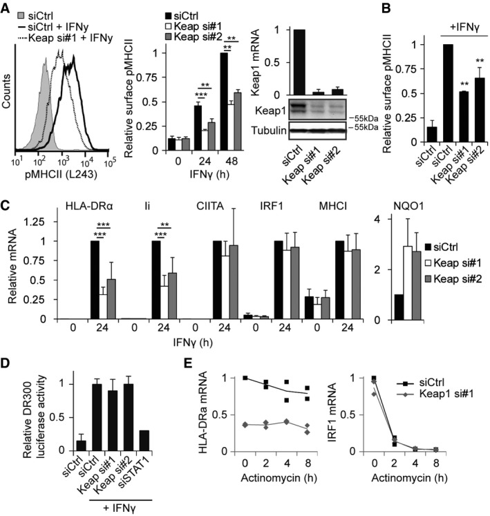
HeLa cells were transfected with siCtrl or siRNAs targeting Keap1 and stained 72 h later for peptide‐loaded MHCII (L243‐cy5), stimulated or not with 100 ng/ml IFNγ for the indicated time. Representative histogram and quantifications are shown. Right: Keap1 silencing was determined by Western blot analysis (bottom) and qRT–PCR (top, normalized to GAPDH).
U118 cells were analysed for MHCII levels according to the same protocol as in (A).
HeLa cells transfected with siCtrl or siKeap1 were either or not exposed to IFNγ for 24 h, and mRNA expression of the indicated genes was analysed using qRT–PCR.
MHCII promoter activity was analysed using a luciferase under control of the MHCII promoter (DR300) in cells transfected with the indicated siRNAs and treated with IFNγ when indicated. siSTAT1 was used as a positive control, and signals were normalized to a Renilla control plasmid.
Cells transfected with siCtrl or siKeap1 were treated with IFNγ for 24 h and lysed, or actinomycin D (2 μM) was added and cells were lysed 2, 4 or 8 h later. mRNA expression level of HLA‐DRα and IRF1 was analysed using qRT–PCR, and IRF1 was used as a control for effectivity of actinomycin D. Individual data points are represented by dots, and the line is the average of the two experiments.
Data information: Experiments shown represent mean + SD of at least three independent experiments (except E,
n = 2). Statistical significance was calculated as compared to control cells using a Student's
t‐test (*
P < 0.05, **
P < 0.01, ***
P < 0.001).
Source data are available online for this figure.

