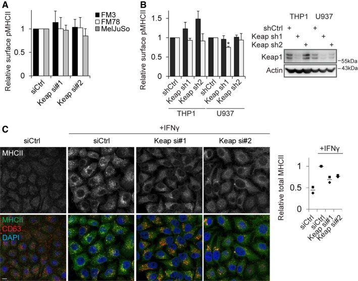
FM3, FM78 and MelJuSo melanoma cells were transfected with siCtrl or siRNAs targeting Keap1 and stained 72 h later for peptide‐loaded MHCII (L243‐cy5) before analysis by flow cytometry.
THP‐1 and U937 cells stably transfected with the indicated shRNAs were stimulated or not with IFNγ for 48 h, and surface expression of peptide‐loaded MHCII was assessed using flow cytometry. For stimulated samples, the signal for the unstimulated cells was subtracted to allow analysis of the IFNγ‐induced expression. Right: Keap1 silencing was determined by Western blot analysis.
HeLa cells transfected with the indicated siRNAs were stimulated or not for 24 h with IFNγ and stained for MHCII, CD63 (late endosomal marker) and DAPI (DNA). For quantification, average signal intensity of at least six fields was quantified over two independent experiments. Individual experimental averages are represented by dots, and the line is the average of the two experiments.
Data information: Experiments shown in (A and B) represent mean + SD of at least three independent experiments, for (C)
n = 2. Statistical significance was calculated compared to control cells using a Student's
t‐test (*
P < 0.05).
Source data are available online for this figure.

