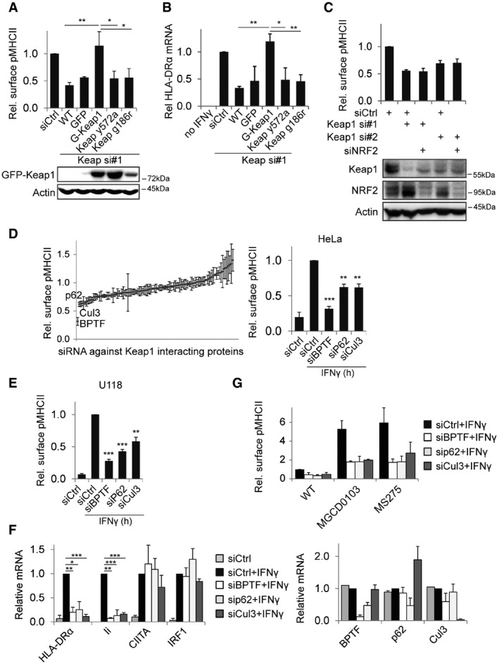
HeLa cells stably expressing GFP or RNAi resistant GFP‐Keap1 with the indicated mutations were transfected with siRNAs and stimulated with IFNγ for 48 h before analysis by flow cytometry. Shown is MFI relative to siCtrl. Bottom panel: Western blot for expression of the indicated GFP‐Keap1 constructs.
HeLa cells as in (A) were stimulated for 24 h with IFNγ, and mRNA levels of HLA‐DRα were measured using qRT–PCR and related to siCtrl.
MHCII expression on HeLa cells transfected with the indicated siRNAs and stimulated with IFNγ for 48 h was measured using flow cytometry and related to siCtrl. Bottom: Western blot analyses for expression of the indicated proteins.
Screen for effect of silencing Keap1 interacting proteins on MHCII surface levels. HeLa cells transfected with 106 different siRNAs targeting Keap1‐interacting proteins were stimulated with IFNγ for 48 h and analysed by flow cytometry. Right: summary of screening data for the indicated proteins.
U118 cells were transfected with the indicated siRNAs and the next day stimulated with IFNγ. 48 h later, MHCII expression was analysed by flow cytometry.
HeLa cells transfected with the indicated siRNAs were stimulated for 24 h with IFNγ and mRNA transcript levels were quantified using qRT–PCR, signal was normalized to GAPDH and siCtrl + IFNγ for each sample. Right graph: knockdown efficiency of the different siRNAs.
HeLa cells transfected with the indicated siRNAs were stimulated for 48 h with IFNγ and the indicated HDAC inhibitors, followed by MHCII expression by flow cytometry.
Data information: All experiments shown represent mean + SD of at least three independent experiments. Statistical significance was calculated compared to control cells using a Student's
< 0.001).

