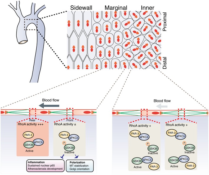
Figure 7. Discrete regulation of axial cell polarity and inflammation by the balance between the PAR‐3/aPKCλ and aPKCλ/GSK3β complexes.

Laminar flow discretely controls endothelial polarity toward the flow axis and vascular inflammatory responses. In the aortic arch, EC Golgi at the sidewall and marginal regions are polarized toward the flow direction. On the other hand, ECs at the inner curvature are not polarized toward the flow direction and have a high risk for atherosclerotic plaque formation. ECs at the marginal region and the inner curvature in Pard3 iΔEC mice showed misoriented Golgi, while those at the sidewall do not. Middle flow restricted RhoA activation to the rear region of ECs, resulting in the non‐uniform distribution of the PAR‐3/aPKCλ and aPKCλ/GSK3β complexes along the flow direction. In the front region, aPKCλ forms a complex with PAR‐3, and hence, the free inactive form of GSK3β is increased. This GSK3β inactivation mediates both EC planar cell polarization and pro‐inflammatory responses. While spatial distribution of GSK3β activation would be important of EC polarity toward the flow axis, local activation of GSK3β is not essential for vascular inflammation.
