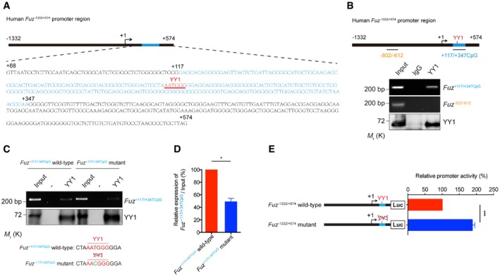
Figure 4. YY1 interacts with Fuz promoter in vivo and in vitro .

-
ASchematic representation of the human Fuz −1332/+574 promoter region. The Fuz +68/+574 was enlarged to show the detailed nucleotide sequence. A putative CpG island was found within Fuz +68/+574 and was defined as Fuz +117/+347CpG. “+1” is the transcriptional initiation site (arrow). The CpG island is highlighted in blue, and sequence of the YY1 binding site is underlined and highlighted in red.
-
BChromatin immunoprecipitation assay demonstrated the binding between YY1 protein and Fuz +117/+347CpG DNA fragment. The blue bar indicates the Fuz +117/+347CpG sequence. However, Fuz −802/−612 (orange) represents a region that did not show interaction with YY1 protein. n = 3.
-
C, D(C) In vitro DNA binding assay demonstrated the binding between purified YY1 protein and purified Fuz +117/+347CpG DNA fragment. When a mutation (green) was introduced to the YY1 site (red) within the Fuz +117/+347CpG fragment, YY1 binding was reduced. (D) is the quantification of the intensity of the Fuz +117/+347CpG DNA bands as shown in (C). Error bars represent s.e.m., n = 3. Statistical analysis was performed using two‐tailed unpaired Student's t‐test. *P < 0.05.
-
EMutating the YY1 binding site in the Fuz promoter increased its transcriptional activity in HEK293 cells. Error bars represent s.e.m., n = 5. Statistical analysis was performed using two‐tailed unpaired Student's t‐test. ***P < 0.001.
