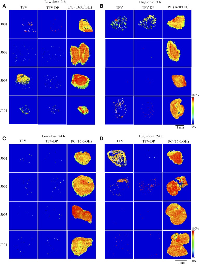
Fig. 4.
Time and dose dependence of TFV and TFV-DP distribution in human colorectal tissue. Representative MALDI MS ion images of (left) TFV, (middle) TFV-DP, and (right) PC(16:0/OH) in colorectal biopsy sections of four different research participants, J001, J002, J003, and J004 collected (A) 3 hours following low-dose, (B) 3 hours following high-dose, (C) 24 hours following low-dose, and (D) 24 hours following high-dose TFV- containing enema treatment. Spatial resolution for MALDI MS images, 50 μm. Scale bar, 1 mm.

