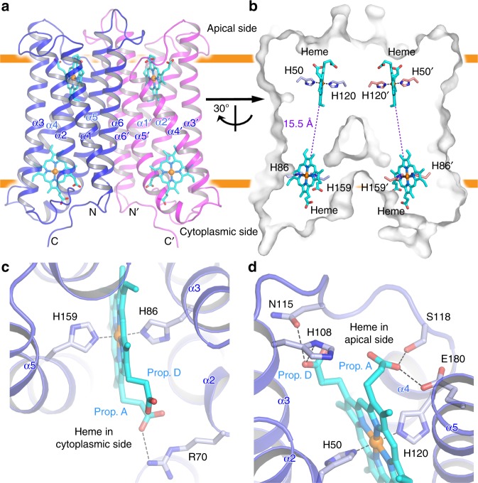
Fig. 1.
Overall structure of human Dcytb. a Ribbon representation of the Dcytb homodimer. Each monomer consists of six α-helices (α1−α6). Both N- and C- termini are located on the cytoplasmic side. Each monomer contains two heme b molecules, coordinated by four highly conserved His residues in a six-coordinate low spin form. b The edge-to-edge distance between two heme molecules is 15.5 Å. c The environment of the heme bound to the cytoplasmic side of Dcytb is shown. d The environment of the heme bound to the apical side of Dcytb is shown

