Fig. 4.
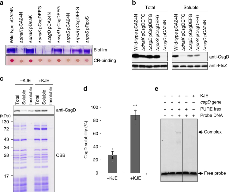
DnaK contributes to CsgD folding. a Biofilm formation and curli production by indicated strains were analysed as shown in Fig. 1. b Protein folding states of CsgD were analysed by immunoblotting. FtsZ served as a control. c CsgD was synthesized in a cell-free translation system in the absence and presence of DnaK-DnaJ-GrpE (KJE). Proteins were separated into soluble and insoluble fractions by centrifugation and CsgD was detected by immunoblotting. Molecular masses are indicated to the left of the panel. d Solubility of CsgD was quantified based on the intensity of protein bands shown in panel c. Experiments were repeated at least three times and average values with standard errors and data plots are shown. **P < 0.01. e DNA-binding activity of CsgD generated in the cell-free translation system was examined by gel-shift assay. The double-stranded DNA fragment harbouring the csgB promoter region was probed with Alexa 488 and incubated with the indicated reaction mixtures. +, presence; −, absence. Full-size scans of immunoblots are shown in Supplementary Figs. 8 and 9

