Fig. 3.
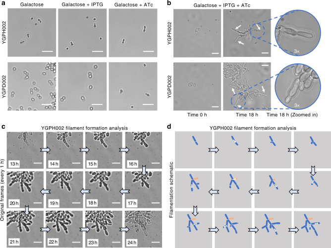
Implementing inducible synthetic regulatory networks for pseudohyphal growth induction. a Inverted microscope images of haploid YGPH002 and diploid YGPD002 strains in different induction conditions. Images were taken after 24 h of growth in galactose media or galactose media supplemented with either 1 mM IPTG or 250 ng/ml ATc. Brightfield images were acquired using a 20× objective. Small white lines are scales which correspond to 20 μm. b Images of the haploid YGPH002 and diploid YGPD002 strains taken at time 0 h or after 18 h of growth inside the ONIX microfluidic platform in galactose media with 250 ng/ml of ATc and 1 mM IPTG supplemented. Areas of the 18 h time point frames have been enlarged by 3× to highlight filament formation. Brightfield images were acquired using a 60× objective. White arrows are pointing to newly formed filaments. Small white lines are scales which correspond to 10 μm. c Time-lapse microscopy and (d) filament formation analysis of the haploid YGPH002 strain when pseudohyphal growth is induced. Cells were induced for 24 h. Here, frames from 1 h intervals starting from the 13 h time point were selected and magnified in order to highlight the generation of branched formations from cells following a unipolar pattern. White arrows highlight growth of new branches from mother cells. Brightfield images were taken using a 60× objective. The white lines correspond to scales with a length of 10 μm. The cell shown in orange is not clearly visible after the 8th frame since it’s covered by other cells

