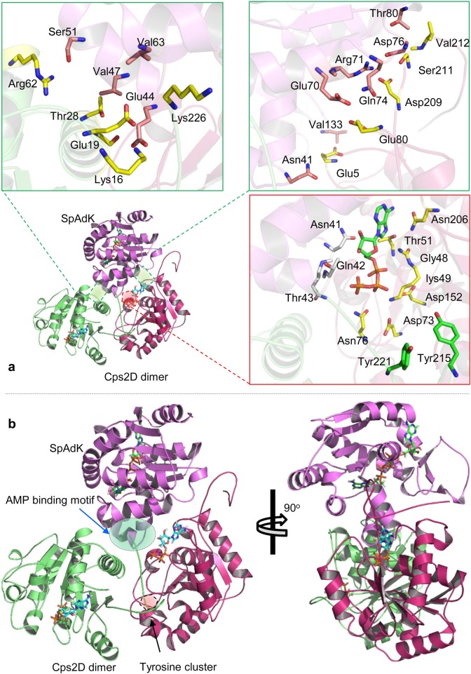
Fig. 7. The binding mechanism of Cps2D and SpAdK.
a The binding interface of the trimeric (Cps2D)2SpAdK. Green boxes highlight the protein interface of SpAdK with each monomeric Cps2D. The red boxes illustrate the role of ATP and the ATP-binding interface in the Cps2D–SpAdK interaction and the autophosphorylation mechanism. b Front and lateral views of the (Cps2D)2SpAdK complex. The red shaded area highlights the C-terminus tyrosine cluster bound to the active site of Cps2D, whereas the green shaded area illustrates the “V”-shaped AMP-binding motif of SpAdK anchoring oligomeric Cps2D

