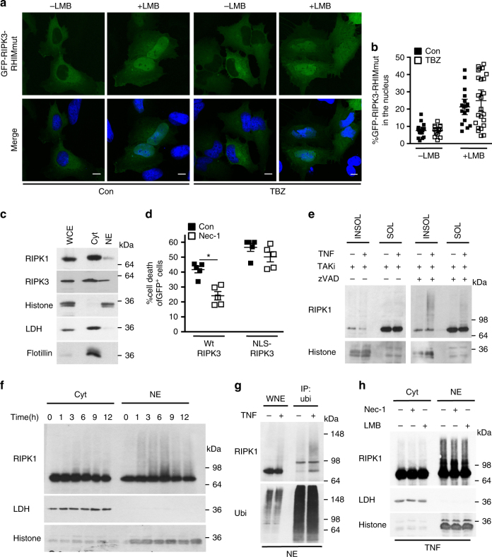
Fig. 2. Nuclear RIPK1 is ubiquitinated during necroptosis.
a Confocal images of single-optical sections of HeLa cells expressing GFP-RIPK3 RHIMmut treated with control (con) or TNF, BV6 and zVAD-fmk (TBZ), with or without LMB. Scale bars: 10 µm. b Quantification of the percentage of nuclear GFP-RIPK3-RHIMmut transiently expressed in HeLa cells and treated with control (con) or with TBZ, with or without LMB. 18–25 transfected cells of a representative experiment of three independent experiments were analyzed. Plots indicate averages ± S.E.M. c Immunoblot of intracellular distribution of RIPK1 and RIPK3 in FADD-deficient Jurkat cells comparing actual protein levels. Lactate dehydrogenase (LDH): cytosolic marker; Histone: nucleus; Flotillin: lipid rafts. Cyt: cytosolic fraction; NE: nucleus-enriched fraction; WCE: whole-cell extract (10%). d Cell death profile of HeLa cell expressing either GFP-RIPK3 or GFP-NLS-RIPK3 treated with TBZ with or without Nec-1 analyzed by SYTOX Blue uptake in the GFP+ population. n = 5; *p < 0.01. e Immunoblots of RIPK1 in the 1% NP-40-soluble and -insoluble fractions of wild-type MEF cells treated with TT (TNF and TAKi) or TTZ for 4 h. f Immunoblot of RIPK1 in cytoplasmic and nuclear fractions isolated from FADD-deficient Jurkat cells treated for the indicated times with TNF. RIPK1 levels were equalized between nuclear and cytosolic fractions by loading four times more of the nucleus-enriched fraction. g Immunoblot of RIP1 in anti-ubiquitin immunoprecipitation of NE of FADD-deficient Jurkat cells treated with TNF for 3 h. WNE: whole-nuclear extract. h Immunoblot of the relative amounts of cytoplasmic and nuclear RIPK1 of FADD-deficient Jurkat cells pre-treated with Nec-1 or LMB and then with TNF (3 h). All immunoblots are representative of two or three independent experiments. Uncropped images of immunoblots are shown in Supplementary Fig. 15

