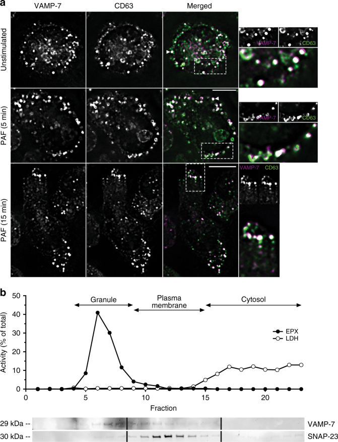
Fig. 1.
VAMP-7 expression and co-localization with crystalloid granules. a Unstimulated and PAF-stimulated (5 μM; 5 and 15 min) adherent eosinophils isolated from the peripheral blood of I5 mice were fixed and stained for VAMP-7 and CD63 immunoreactivity. Arrowheads (white) indicate co-localization of VAMP-7 and CD63 to the membranes of crystalloid granules. Stimulated cells represented 30–40% of total population in 10 high-powered fields. Scale bar for merged PAF (5 min) applies to top and middle panels, 5 μm; scale bar for merged PAF (15 min) applies to lower panel only, 5 μm. Images at right are magnified from boxes shown in dotted lines. White indicates magenta-green overlap. b Subcellular fractionation of eosinophils (5 × 10⁷) isolated from peripheral blood of four I5 mice followed by western blot analysis of fractions. In subcellular fractions, EPX was detected in fractions 5–9, and lactate dehydrogenase+ cytosolic fractions were enriched in fractions 15–23. Western blot analysis showed VAMP-7 concentrated in fractions 5–10, while SNAP-23 (cognate Q-SNARE for VAMP-7) was found in fractions 10–14. Representative of three independent experiments

