Fig. 6.
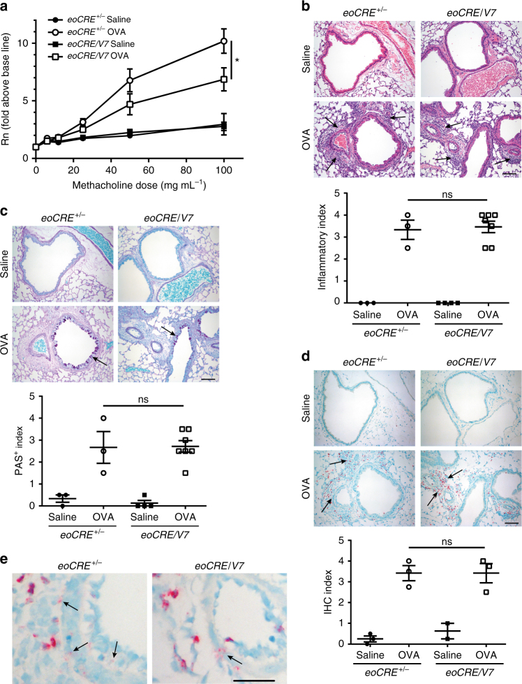
Cell-specific gene deletion of VAMP-7 in eosinophils leads to reduced airway hyperresponsiveness. a eoCRE+/− and eoCRE/V7 mice (8 weeks old) were subjected to acute OVA or saline (control) treatment, and subsequently challenged with aerosolized methacholine. Airway resistance (Rn) presented as fold above baseline was assessed using an invasive ventilator-based technique (Flexivent). Values are mean ± SEM. All measurements, n = 13. *p < 0.05 using ANOVA with Tukey’s post hoc analysis. b Representative H&E histological images of lung sections from eoCRE+/− and eoCRE/V7 mice subjected to saline or OVA treatment. Arrows indicate regions of inflammation. c Assessment of goblet cell metaplasia and airway epithelial cell mucin accumulation by PAS staining of adjacent lung sections to those shown in (b). Arrows indicate mucin-producing goblet cells in the large airways of the lungs. d Immunohistochemical staining of similar lung sections shown in (b) and (c) with anti-MBP. Arrows indicate regions of MBP+ eosinophilic infiltration. Scale bars, 50 μm. Graphs below each panel of images show quantitative histology, carried out in a randomized, blinded, and unbiased analysis. Data are presented as mean ± SEM of sections of lung measuring ≥200 mm2 (3–7 mice per condition). e Higher magnification of lung sections stained with anti-MBP from (d) to indicate extracellular MBP release. Scale bar, 20 μm. Arrows indicate regions of MBP extracellular deposition resulting from non-cell associated eosinophil degranulation

