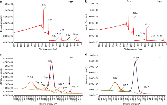
Fig. 3.
XPS of the implant surfaces. Survey spectra of a the TiMA and b TiNT surfaces. The two spectra appeared very similar in elemental composition indicating no notable surface contamination due to the multiple processing steps required for the TiNT surface. c, d Deconvolution of the Ti envelop for c TiMA surface and d TiNT surface. TiMA surface had a thin (<10 nm) TiO2 layer as the underlying metal peak is seen at 453.2 eV (arrow). On the contrary, this and the sub-oxide peaks at <456 eV were absent in the TiNT surface, on which the oxide layer was >10 nm (the sampling depth of the technique)

