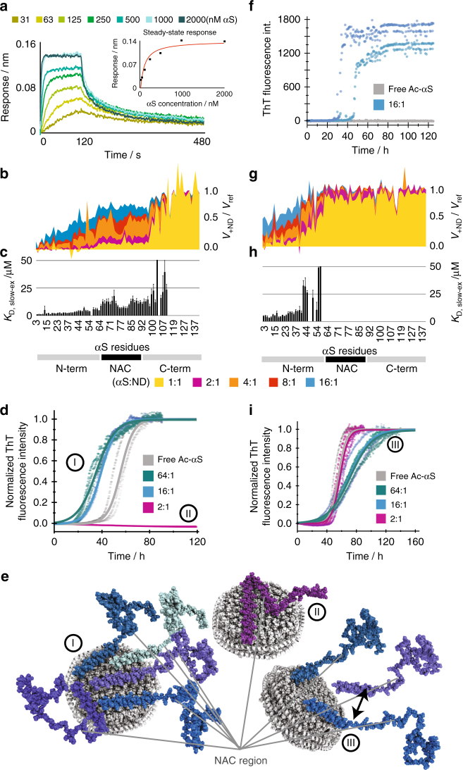
Fig. 5.
The interplay between interaction kinetics, differential residue specific affinities, membrane charge density and accessible surface area modulates αS aggregation. a BLI sensorgrams obtained with immobilized 100% POPG NDs and addition of different concentrations of αS. Corresponding steady-state response plot are shown as insert. A fitted global affinity (KD) of 67 ± 17 nM and a fitted off-rate of 0.015 ± 0.006 s−1 could be extracted. b NMR attenuation profiles of a titration of 50 µM αS with varying concentrations of 100% POPG NDs (αS-to-NDs molar ratios ranging from 16:1 to 1:1, see color code). c Corresponding residue-specific affinities extracted from NMR titration data. The values report on the slow-exchange (lower) limit for the affinities (see text for details). d Normalized ThT fluorescence aggregation curves for selected αS-to-ND ratios (conditions identical as in b–g). e NMR-derived binding modes and their link to the indicated aggregation behaviour (see Supplementary Note 3 for more details on how binding models were generated). Although high amounts of NDs with high charge density inhibit aggregation (binding mode II), limited amounts of highly charged membrane surfaces enhances aggregation (binding mode I). For NDs with a moderate lipid charged density, only one αS-binding mode was observed that has little effect on aggregation (binding mode III). f Nucleation ThT assays in quiescent conditions at pH 5.3. Although under these conditions no aggregation is observed in the absence of NDs (duplicates in gray), the presence of 16:1 molar ratio of 100% POPG NDs (duplicates in light and dark blue) induces primary nucleation. g–i Same as data shown in b–d but using NDs with 50% POPG content

