Fig. 5.
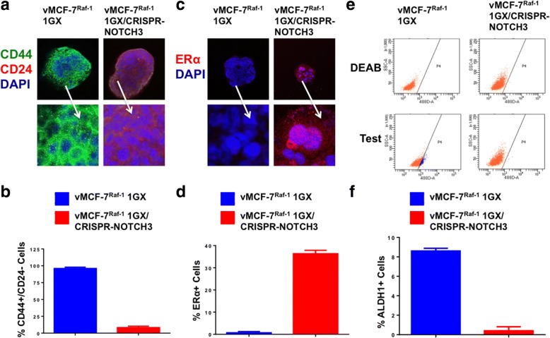
Analysis of breast cancer stemlike phenotype in vMCF-7∆Raf1 1GX cells with abrogated NOTCH3 expression. a Immunofluorescence analysis showing representative images of vMCF-7∆Raf1 1GX and vMCF-7∆Raf1 1GX/CRISPR-NOTCH3 cells stained in green with a CD44 polyclonal antibody and in red with a CD24 monoclonal antibody. Nuclei were stained in blue with 4′,6-diamidino-2-phenylindole (DAPI). b Graph showing the average of cells expressing a CD44+/CD24− phenotype from three independent experiments (± SD). c Immunofluorescence analysis showing representative images of vMCF-7∆Raf1 1GX and vMCF-7∆Raf1 1GX/CRISPR-NOTCH3 cells stained in red with an estrogen receptor alpha (ERα) monoclonal antibody. Nuclei were stained in blue with DAPI. d Graph showing the average of ERα-positive cells from three independent experiments (± SD). e Fluorescence-activated cell sorting analysis showing aldehyde dehydrogenase 1 (ALDH1) activity in vMCF-7∆Raf1 1GX and vMCF-7∆Raf1 1GX/CRISPR-NOTCH3 cells. Samples treated with the ALDEOFLUOR inhibitor N,N-diethylaminobenzaldehyde (DEAB) were used as a negative control. f Graph showing the average of ALDH1+ cells from three independent experiments (± SD)

