Fig. 7.
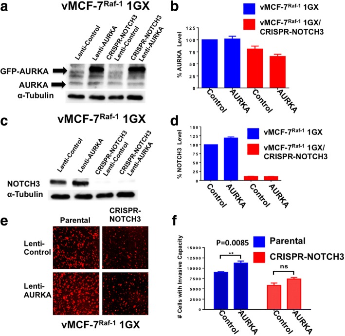
Invasive capacity of vMCF-7∆Raf1 1GX cells expressing a green fluorescent protein (GFP)-tagged kinase Aurora kinase A (AURKA) construct. a Immunoblot assay showing expression of endogenous and GFP-tagged AURKA in vMCF-7∆Raf1 1GX and vMCF-7∆Raf1 1GX/CRISPR-NOTCH3 cells. b Densitometric analysis showing the percentage of endogenous AURKA protein levels in vMCF-7∆Raf1 1GX and vMCF-7∆Raf1 1GX/CRISPR-NOTCH3 cells relative to control. Graph shows the average from three independent experiments (± SD). c Immunoblot assay showing NOTCH3 protein levels in vMCF-7∆Raf1 1GX and vMCF-7∆Raf1 1GX/CRISPR-NOTCH3 cells expressing empty lentiviral vectors (control) and lentiviral GFP-tagged AURKA vectors. d Densitometric analysis showing the percentage of NOTCH3 protein levels in vMCF-7∆Raf1 1GX and vMCF-7∆Raf1 1GX/shRNA-NOTCH3 cells relative to vMCF-7∆Raf1 1GX cells infected with empty lentiviral vectors (control). Graph shows the average from three independent experiments (± SD). e In vitro real-time invasion assay of vMCF-7∆Raf1 1GX and vMCF-7∆Raf1 1GX/CRISPR-NOTCH3 cells expressing empty lentiviral vectors (control) and lentiviral GFP-tagged AURKA vectors. f Graph showing the average number of invasive cells from three independent experiments (± SD)

