Figure 1.

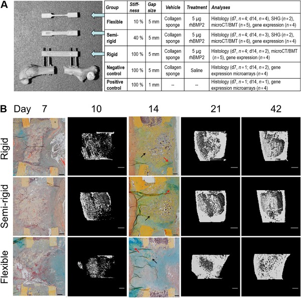
(A) Animal model. Defect model and sample overview. Rat femur with a 5‐mm defect gap and attached external fixator (RatExFix, RISystem) of rigid stability, accompanied by fixator bars of the semi‐rigid and flexible stabilities. Numbers of animals used for each analysis are given. SHG = second‐harmonic generation imaging; BMT = biomechanical testing. (B) Course of defect healing. Healing was similar in all groups at days 7 and 10. By day 14, the rigid and semi‐rigid group showed more woven bone (black arrows) at the defect site with islands of cartilage (red arrows). In contrast, at day 14, the flexible group showed less woven bone, with more proliferating cells across the gap and huge cartilage islands at periosteal site opposite to the fixator location. By day 21 and day 42, respectively, all groups revealed bony defect bridging (histology: Movat Pentachrome staining, scale bars = 500 μm; μCT: scale bars = 1 mm).
